


 shine@shieldconnect.in
Neurodegenerative Disorders:
Neurodegenerative disorders are conditions that involve cell death and degeneration over time. Examples are Alzheimer’s, and Parkinson’s.
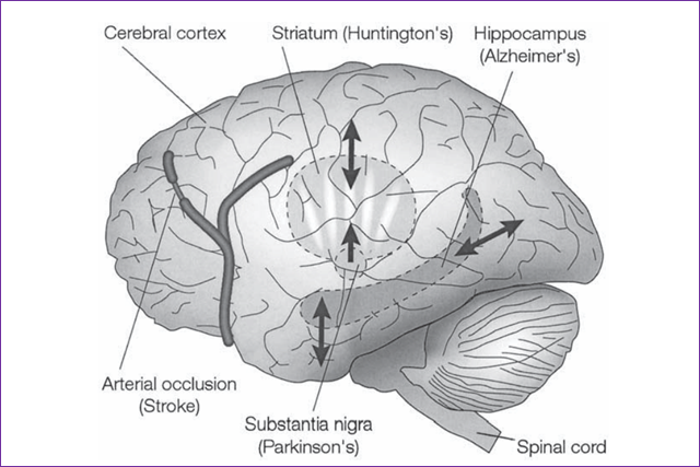
The cerebral cortex belongs to Cerebellum and is the main area involving thinking, decision-making, emotions, and the five senses.
Hippocampus:
The primary role is memory formation classifying the information and long-term memory. People with extensive damage to this part of the brain may suffer amnesia.
Stratum:
The striatum is the input module to the basal ganglia, a neuronal circuit necessary for voluntary movement control.
Different populations of neurons are selectively vulnerable to different
neurodegenerative disorders.
In AD, neurons in the hippocampus and certain regions of the cerebral cortex degenerate.
In PD, it is the dopaminergic neurons in the substantia nigra that undergo apoptosis
In Huntington’s disease (HD), it is neurons in the striatum that die; and
Which neurons die in stroke depends on which blood vessel is affected, but often it is neurons in the cerebral cortex and striatum.
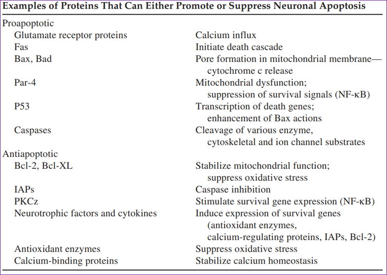
Although the genetic and environmental factors that trigger neuronal apoptosis may be different in physiological and pathological settings, many of the subsequent biochemical events that execute the cell death process are highly conserved.
One key focus of this death program is the mitochondrion, which controls the decision of cell death. Changes that occur in mitochondria in cells undergoing apoptosis include increased oxyradical production, the opening of pores in their membranes, and the release of cytochrome c. These mitochondrial changes are central to the cell-death process because agents such as manganese superoxide dismutase and cyclosporine A, which act
directly on mitochondria to suppress oxidative stress and membrane pore formation, also prevent neuronal death in various experimental models.
ANTI-APOPTOTIC SIGNALING:
There are several prominent anti-apoptotic signaling pathways in neurons. Activation of neurotrophic factor receptors can protect neurons against apoptosis by activating receptors linked via kinase cascades to the production of cell-survival-promoting proteins. Brain-derived neurotrophic factor (BDNF), nerve growth factor (NGF), and basic fibroblast growth factor (bFGF) can prevent the death of cultured neurons, in part by stimulating the production of antioxidant enzymes, Bcl-2 family members, and proteins involved in the regulation of Ca2+ homeostasis. Tumor necrosis factor-(TNF-_), ciliary neurotrophic factor (CNTF), and leukemia inhibitory factor (LIF) are three
cytokines that can prevent neuronal death in experimental models of natural neuronal death and neurodegenerative disorders.
Alzheimer’s Disease: Progressive impairment of cognition and emotional disturbances characterize AD; these symptoms result from degeneration of synapses and death of neurons in limbic structures such as the hippocampus and amygdala, and associated regions of the cerebral cortex. The damaged neurons exhibit aggregates of hyperphosphorylated tau protein and evidence of excess
Parkinson’s Disease: A Motor System Disorder in which Patients with PD exhibit profound motor dysfunction as the result of degeneration of dopaminergic neurons in their substantia nigra. The cause(s) of PD is unknown, but likely involves increased oxidative stress and mitochondrial dysfunction in dopaminergic neurons.
Ischemic Stroke: Ischemic brain damage resulting from occlusion of a cerebral blood vessel is characterized by an infarct with a necrotic core in which all cells die rapidly and a surrounding ischemic penumbra in which neurons die over days to weeks.
Metabolic compromise, overactivation of glutamate receptors, Ca2+ overload, and increased oxyradical production occurs in neurons subjected to ischemia.
In addition, complex cytokine cascades involving microglial cells and the cerebrovasculature may play important roles in promoting or preventing neuronal death after stroke
Traumatic Brain and Spinal-Cord Injury
The leading cause of death and disability in persons under the age of 40 is a traumatic injury to the brain and spinal cord.
Trauma initiates biochemical and molecular events involving many of the same neurodegenerative cascades and neuroprotective signalling mechanisms that occur in the chronic neurodegenerative diseases described above.
Studies have documented apoptosis-related changes in neurons including the presence of DNA strand breaks, caspase activation, and increased Bax and p53 expression.
NGF infusion beginning 24 h after TBI results in improved learning and memory and decreased death of neurons
Schizophrenia: Schizophrenia is a serious mental disorder in which people interpret reality abnormally. Symptom groups in schizophrenia include,
Positive symptoms: Positive symptoms include delusions and hallucinations, linked to aberrant salience. These symptoms are most recognizable during periods of acute psychosis.
● Cognitive symptoms: Impairments in learning, memory, attention, and executive functioning are all included as cognitive symptoms.
● Negative symptoms: Negative symptoms include blunting of affect (lacking emotional expression), avolition (deficits in motivation), and social withdrawal.
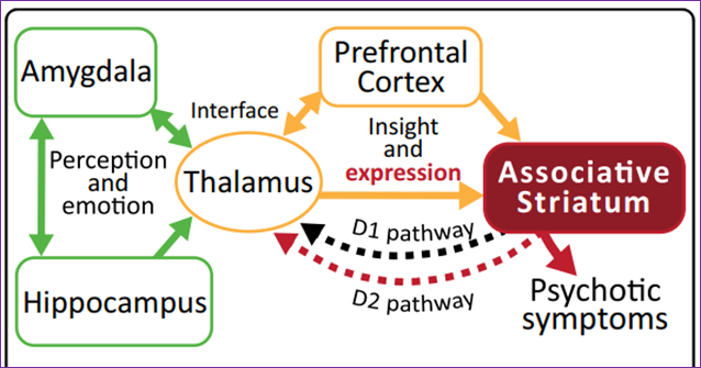
Dysfunction in a variety of brain regions can elicit psychotic symptoms.
A primary circuit involved in psychosis includes the thalamus and prefrontal cortex (yellow) feeding into the associative striatum.
Alterations in the thalamus and prefrontal cortex are involved in hallucinations and insight for delusional symptoms.
Expression of psychotic symptoms in most cases requires increased activity in the associative striatum and specifically excessive D2 receptor stimulation (red).
Other limbic regions such as the hippocampus and amygdala (green) can feed into this circuit contributing to altered sensory perception and emotional context