


 shine@shieldconnect.in
Facts about Brain
Brain Structure/Anatomy and Function
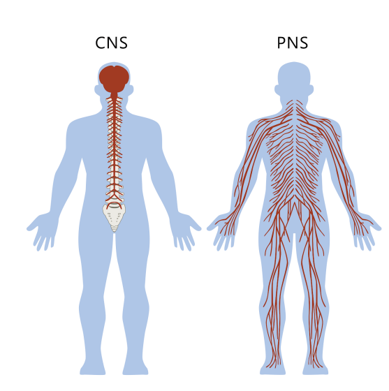
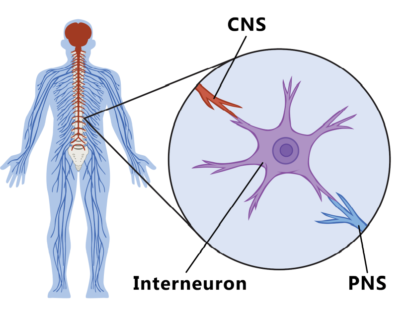
Central Nervous system (CNS)
Made up of the brain and spinal cord which controls the regulation of body systems and the Processing and creation of memories.
Peripheral Nervous System (PNS)
Made up of all the neurons connecting the CNS to the rest of the body.
The PNS sends messages from the body to the spinal cord and/or brain & back. The PNS is divided into two sections:
Autonomic Nervous System (ANS): Unconscious Control of the body
Somatic Nervous System (SNS): Conscious Control of the body.
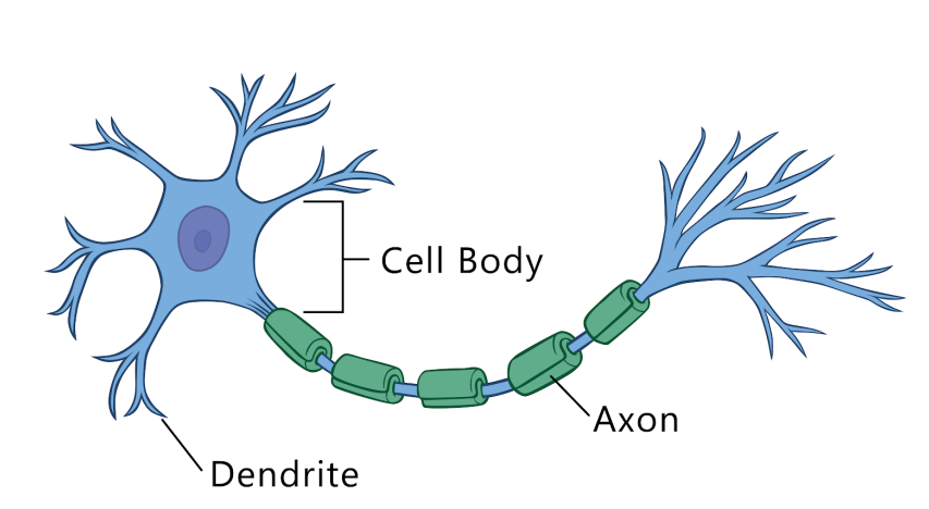
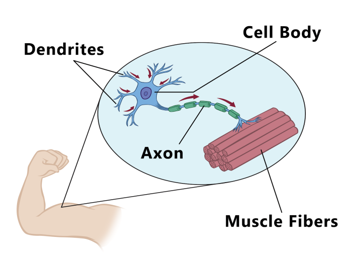
A cell is the basic unit of life: Similarly, a neuron is the basic structure of the Brain.
Neurons /nerve cells carry signals throughout the nervous system.
Groups of nerve cells get together to form the nerve tissue or nerves.
A signal can pass through a nerve cell at a speed of 265 miles/hour.
The cell body
constitutes the main part of a nerve cell.
contains the main control center of the cell
Extends several hair-like branches called dendrites (singular: dendrite).
Dendrites
carry signals to the cell body from other neurons or the environment.
Axon:
Long, thick, tail-like structure attached to the cell body called the axon
Carry the signals away from the cell body.
The dendrites and axons are called nerve fibers
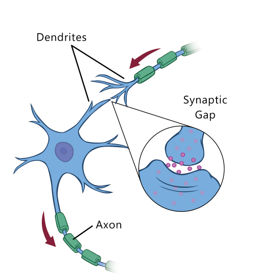
A neuron receives a signal through the dendrites,
which is then transmitted to the cell body and then through the axon.
Once at the end of the axon the signal will be transmitted to the next neuron.
No two neurons touch each other
Myelin sheath:
A fatty layer acts as a layer of insulation.
Prevents the nerve signals of one neuron from interfering with another neuron.
Surrounds all the dendrites & the axon.
The nerve cells of the PNS have another protective covering on top of the
myelin sheath known as neurilemma which is made up of living cells.
The neurilemma helps damaged nerve fibers regrow and recover.
Many of the nerve cells of the brain & the spinal cord don’t have a myelin sheath nor a neurilemma.
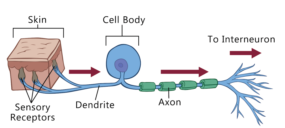
Classification of Neurons
Sensory neurons: Signals from the body to the brain /SC
Interneurons: connect the sensory neurons with the motor neurons.
Motor neurons: Signals from the brain and/or the spinal cord to the body
FACTS about Neurons
Many of the cells in our body can grow and multiply as the body grows. Nerve cells cannot be replaced.
A baby is born with over one trillion neurons. As the baby grows, so do the neurons.
The dendrites and axons become longer and longer making new connections with other neurons.
After a person has reached adulthood, some nerve cells begin to die.
Since neurons cannot be replaced, the number of nerve cells in the body begins to reduce.
Elderly persons tend to forget information often or are unable to move parts of their body like as used to when they were younger.
A neuron is a basic structure
Neurons /nerve cells carry signals throughout the nervous system.
Groups of nerve cells get together to form the nerve tissue or nerves.
A signal can pass through a nerve cell at a speed of 265 miles/hour.
The cell body.
constitutes the main part of a nerve cell.
contains the main control center of the cell
Extends several hair-like branches called dendrites (singular: dendrite).
Dendrites
carry signals to the cell body from other neurons or the environment.
Axon:
Long, thick, tail-like structure attached to the cell body called the axon
Carry the signals away from the cell body.
The dendrites and axons are called nerve fibers.
A neuron receives a signal through the dendrites,
which is then transmitted to the cell body and then through the axon.
Once at the end of the axon the signal will be transmitted to the next neuron.
No two neurons touch each other.
Myelin sheath:
A fatty layer acts as a layer of insulation.
Prevents the nerve signals of one neuron from interfering with another neuron.
Surrounds all the dendrites & the axon.
The nerve cells of the PNS have another protective covering on top of the
myelin sheath known as neurilemma which is made up of living cells.
The neurilemma helps damaged nerve fibers regrow and recover.
Many of the nerve cells of the brain & the spinal cord don’t have a myelin sheath nor a neurilemma.
Neurons are classified into 3 types.
Sensory neurons: Signals from the body to the brain /SC
Interneurons: connect the sensory neurons with the motor neurons.
Motor neurons: Signals from the brain and/or the spinal cord to the body
Many of the cells in our body can grow and multiply as the body grows. Nerve cells cannot be replaced.
A baby is born with over one trillion neurons. As the baby grows, so do the neurons.
The dendrites and axons become longer and longer making new connections with other neurons.
After a person has reached adulthood, some nerve cells begin to die.
Since neurons cannot be replaced, the number of nerve cells in the body begins to reduce.
Elderly persons tend to forget information often or are unable to move parts of their body like as used to when they were younger.
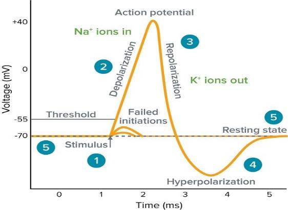
Information Exchange in the Nervous System:
Neurons constantly relay information between each other, and between themselves and their environment
Neurotransmitters are the body’s chemical messengers and transmit messages between neurons, or from neurons to muscles.
Communication between two neurons happens in the synaptic cleft (the small gap between the neurons)
Functions of Different Regional Functions of the Brain:
Hindbrain or Reptilian Brain:
This controls human’s primitive instincts and most basic functions. Consider instincts for survival, dominance, mating, and basic functions of respiration and heartbeat. This contains the Spinal Cord, Medulla Oblongata, Pons, and Cerebellum.
The Limbic System (Emotional Brain)
This is where human emotions reside, where memory begins, and where these two functions combine to make behaviors with positive or negative feelings.
It’s where most unconscious value judgments are made.
Information going through the Limbic system is filed under ‘agreeable’ or disagreeable, and it also plays a role in attention, spontaneity, and creativity.
Located in the Limbic system are the Amygdala, Hippocampus, Hypothalamus, and Thalamus.
Neocortex :
Describe two-thirds of the brain (overlapping numerous smaller regions).
Comprises the Frontal Lobe, the Parietal Lobe, the Temporal Lobe, the Occipital Lobe, Broca’s Area, and the Corpus Callosum.
Brain Pruning:
Little-used areas of the brain’s white matter dissolve during three distinct periods, leaving the brain more efficient and, with each pruning, opening up more abilities.
This occurs repeatedly in infancy, between puberty and mid-adolescence (roughly aged 14 and 17, +/- a year), and again in the early twenties (about age 22, +/- a year).
For instance, the first pruning enables abstract philosophical thought.
Schizophrenia emerges following the third pruning.
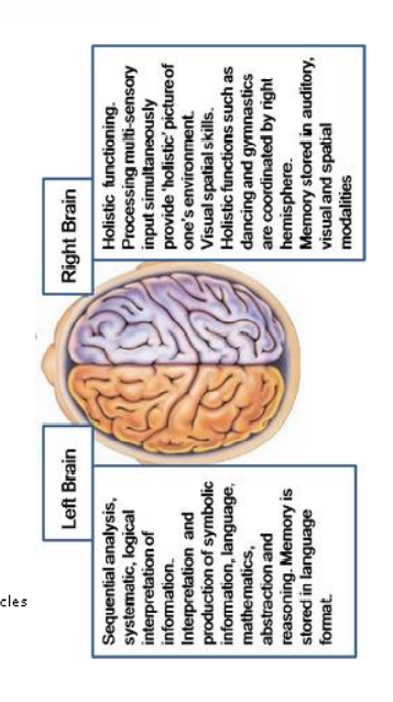
Right and Left Hemispheres of the Brain –
The brain is divided in half, with the Corpus Callosum bridging the two hemispheres.
The right side of the brain controls the left side of the body, and the left side of the brain controls the right side of the body.
The right side of the brain is generally more concerned with the artistic, spatial, and musical inclinations while the left is more orientated toward the colder, linear, rational, and verbal aspects.
The left brain
contains many areas which are vital for the processing and formation of speech. Helps children acquire language, make the connection between sounds heard and things seen or otherwise experienced, and seems to control speech in 96% of children.
The left brain is good at exact and precise thought processes, and the pre-motor areas of this region deal with grammar.
The left side of the brain controls actual speech movements that generate sounds.
The Left Hemisphere of the brain has 186 million more neurons than the Right Hemisphere.
The right-side Brain:
Involved in interpreting and generating speech with meanings.
The right brain is apt at making broad and sweeping understandings and it can also decipher meaning by ‘the way you said it
The point of emphasis and comprehending humor.
People with Dyslexia have a slightly larger Right Hemisphere.
Amygdala :
Helps in storing and classifying emotionally charged memories.
Plays a significant role in producing emotions, especially fear and jealousy.
In teenagers, the Amygdala is more active than the Frontal Lobe;
individuals with an enlarged Amygdala have been diagnosed with manic-depressive disorder.
Broca’s Area :
This part of the Neocortex controls speech, language recognition, and facial nerves.
Brain Plasticity – A process that refers to how nerve cells and neurons physically change inside the brain, in response to a change in environmental circumstances over time and/or in response to brain injury.
Cerebellum –
The portion of the brain (located in the back of the brain) helps coordinate movement, such as balance, posture, movement, and muscle coordination.
The Cerebellum contains half of all the neurons in the brain but comprises only 10% of the brain Cerebral Cortex – This is the main area involving thinking, decision-making, emotions, and the five senses.
Corpus Callosum:
This structure connects the right and left hemispheres of the brain and facilitates communication between the two.
Important for intelligence, consciousness, and self-awareness.
The largest White Matter structure in the brain and reaches full maturity in the twenties.
Frontal Lobe:
The front part of the brain is involved in planning, judgment, reasoning, impulse control, organizing, problem-solving, selective attention, personality, personality, and a variety of “higher cognitive functions”, including behavior and emotions.
The Prefrontal cortex (front of the Frontal Lobe) is important to personality.
The posterior (back) of the Frontal Lobe serves to modify movements Both lobes grow measurably between ages10 and 12 (with girls’ growth spurt generally coming a little earlier than boys’), and then shrink in the twenties as extraneous branches and pruned back into efficient, well-organized circuitry.
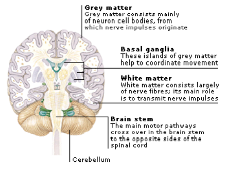
Gray Matter and White Matter –
Gray matter can be thought of as the actual computers themselves, whereas white matter represents the network cables connecting the computers together.
The brain can adapt to white matter damage. Unlike Gray matter, which peaks in development in a person’s twenties, the white matter continues to develop and peaks about middle age.
In terms of oxygen consumed, 6% will be used by the brain’s white matter and 94% by the Gray matter.
The development of Multiple Sclerosis, as well as protein build-up associated with Alzheimer’s both, affect white matter
Hippocampus:
The primary role is memory formation classifying the information and long-term memory.
Leading thought hypothesizes that this area also intuitively informs a person of their place within an environment.
People with extensive damage to this part of the brain may suffer amnesia.
Furthermore, in Alzheimer’s Disease, this area is among the first to suffer damage.
Hypothalamus –
controls many body processes, such as heart rate and feelings of hunger and thirst, as well as circadian (24-hour cycle) rhythms.
located above the brain stem. Failure to get 8-9 hours of sleep each night can disrupt a teen’s circadian rhythm ‘clock’.
Medulla Oblongata:
Governs involuntary processes (breathing, swallowing, defecation, digestion & heart rate).
Located on the lower half of the brain stem, next to the Pituitary Gland.
Occipital Lobe:
Located in the back of the brain which processes visual sensations and information (images, shapes, colours).
The Visual Cortex resides here, and it is where reading is made possible.
Parietal Lobe:
Involved in processing pain and touch sensation, as well as being involved in emotion, memory, and speech.
Integrates auditory, visual, and tactile signals.
Maintains two slightly different functions, depending on the right or left side of the area.
They contain the primary sensory cortex which controls sensation (tough, pleasure).
Behind this is a large association area that controls fine sensation (judgment of texture, weight, size, shape)
Parietal Lobe, right: damage to this can cause visual-spatial difficulties Parietal Lobe, left: damage to this area may disrupt a patients ability to understand spoken and/or written language.
These areas are immature until about the age of 16.
Pituitary Gland:
The pea-sized gland which secretes hormone regulates the body.
Controls temperature regulation, growth, blood pressure, water regulation in the body, breast milk production and sex organ functions of both genders.
The Pons:
Maintains a role in sleep, particularly in terms of being conscious (awake) or not, regulate consciousness and is associated with the sense of higher purpose.
“Locked-in Syndrome”, a condition in which a patient is aware and awake but cannot move or communicate due to complete paralysis except for the eyes, occurs when a lesion damages the Pons.
Temporal Lobe:
Considered that there are two temporal lobes, as one is located about ear-level in each hemisphere of the brain.
They allow a person to distinguish between different smells and sounds. They also help with new information and are believed responsible for short-term memory.
➢ Right-hemisphere lobe: mainly involved in visual memory (memory for pictures, faces)
➢ Left-hemisphere lobe: mainly involved in verbal memory (memory for words, names)
These areas reach their grey matter peak about age 16, followed by a decade of pruning.
Thalamus:
Think of this as the relay station of the brain.
All sensory input and output routes through this point.
It also is suspected to play a function in muscle control.
It is understood is in some manner governs sleep and wakefulness, as well as regulating important bodily functions including hunger, body temperature and breast feeding.
Wernicke’s Area:
Responsible for language recognition.
Located in the Left Hemisphere of 90% of people.
Damage to the Wernicke’s Area affects a person’s ability to string together a coherent sentence or even the loss of the ability to understand language.
Studies suggest the brain’s ability to link letter combinations with sound may not be fully developed until age 11.
Nervous System Disorders
A nervous system that functions correctly is a fantastically complex, well-oiled machine where,
synapses fire appropriately,
muscles move when needed,
memories are formed and stored, and
emotions are well regulated.
Unfortunately, each year millions of people right from infants to the elderly, dealing with some sort of nervous system disorder.
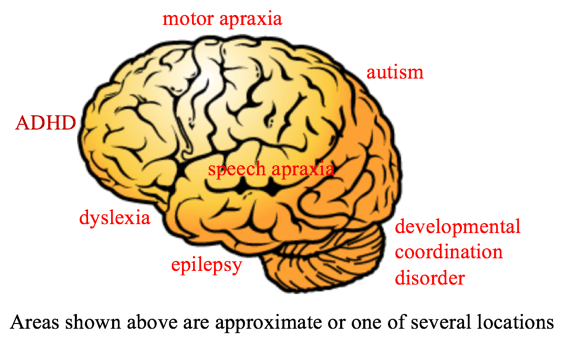
Neurodevelopmental Disorders
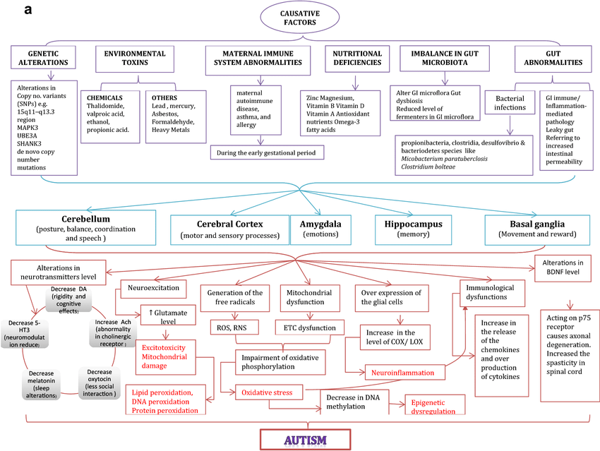
Neurodevelopmental disorders (NDD) are a group of disorders that arise during the developmental period of an individual, before adulthood. This happens due to improper development of the central nervous system, because of interplay between various environmental or genetic factors. Making NDDs as “multifaceted conditions characterized by impairment in cognition, communication, behaviour and/or motor skills resulting from abnormal brain development
The DSM 5, published in 2013, includes the new category of Neurodevelopmental disorders like Pervasive developmental disorder (intellectual disability), ADHD, speech and communication disorders, and Tourette's syndrome are included under NDD.
There is a significant overlap of symptoms between the various NDDs, and they share common characteristics e.g., autism and ADHD, hence using the term NDD helps to acknowledge the similarities and overlap of symptoms between these disorders. Following is the broad classification of NDD in DSM 5:
• Intellectual Disabilities
• Communication disorders (e.g., Speech, language d/o)
• Autism spectrum disorder
• Attention-deficit/hyperactivity disorder
• Specific learning disorder
• Motor disorders (e.g., tic and Tourette’s d/o)
• Other specified/ unspecified neurodevelopmental disorders.
Causes of Neuro-Developmental Disorders:
NDD is an umbrella term that includes many diseases with heterogeneous presentations but the common factor in all these diseases is that they develop because of improper development of the nervous system and occurs during the developmental period of a child before their adulthood.
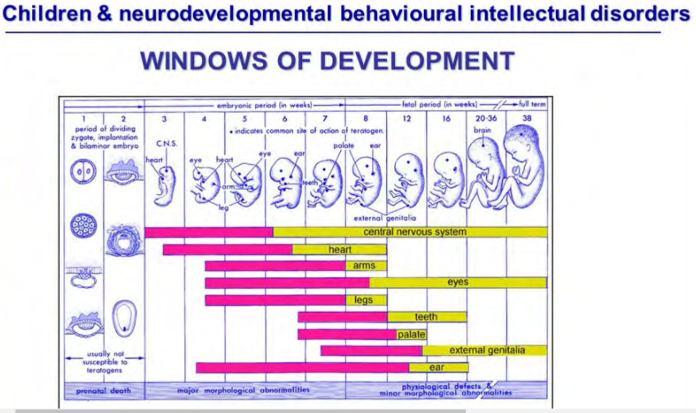
The early developmental period is very sensitive to intrinsic and extrinsic factors which can cause developmental delay.
The most critical periods during the development of a child are
• Preconception period
• Prenatal period
• Postnatal period Factors.
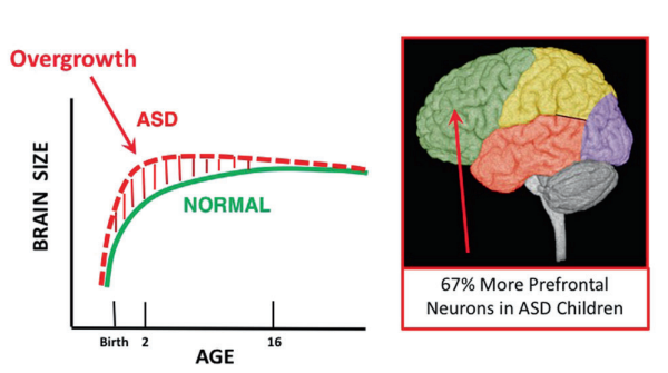
Autism spectrum disorder (ASD):
A neurodevelopmental disorder. Its severity differs from person to person. ASD is four times more prevalent in males than females. A characteristic symptom of ASD includes,
Impaired social skills.
Difficulty making and maintaining eye contact and reading social cues.
Problems feeling empathy for others.
Repetitive motor behaviors, preoccupation with specific subjects, strict adherence to certain rituals, and unusual language use.
30 % ASD develop epilepsy, and Fragile X also have intellectual disability.
Although early interventions can help mitigate the effects of the disease, there is currently no cure for ASD.
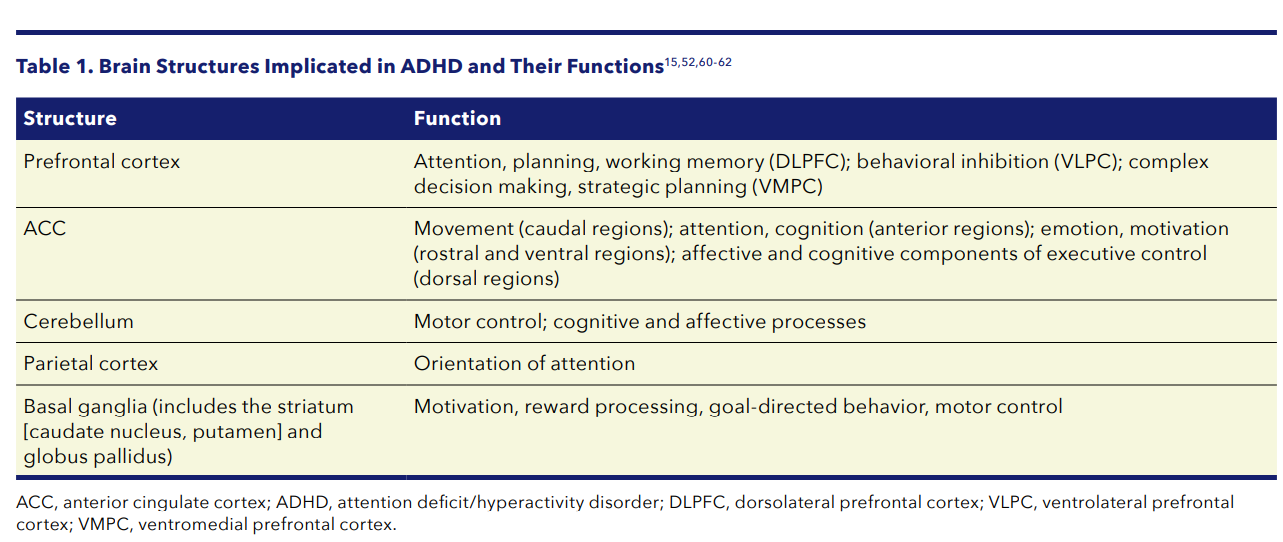
Attention Deficit Hyperactivity Disorder (ADHD)
ADHD is more prevalent in males than females. Symptoms of the disorder include inattention (lack of focus), executive functioning difficulties, impulsivity, and hyperactivity beyond what is characteristic of the normal developmental stage.
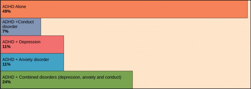
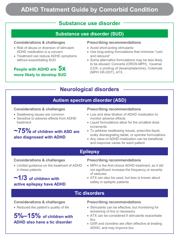
Many people with ADHD also show comorbidity, in that they develop secondary disorders in addition to ADHD. Examples include depression or obsessive-compulsive disorder (OCD).
ADHD and Co-morbid conditions incidence including the following.
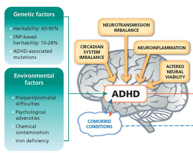
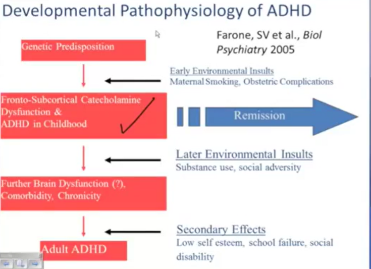
The pathogenesis of ADHD is complex, involving an interplay of genes and the environment
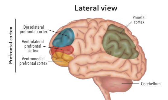
The cause of ADHD is unknown, although research points to a delay and dysfunction in the development of the prefrontal cortex and disturbances in the neurotransmitter imbalance,
neuroinflammation defective
immunoregulation,
circadian system dysfunction & altered neural viability,
neurodegeneration
are pathophysiological processes related to attention-deficit/hyperactivity disorder (ADHD) disorder and their comorbidities.
Environmental factors, including exposure to certain pesticides, may also contribute to the development of ADHD in some patients.
The pathogenesis of ADHD is complex, involving an interplay of genes and the environment. Multiple brain networks are involved in attention, with reduced function of these networks associated with symptoms and characteristics of ADHD, including deficits in sustained attention, response inhibition, executive function, and emotional control
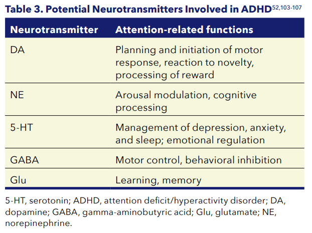
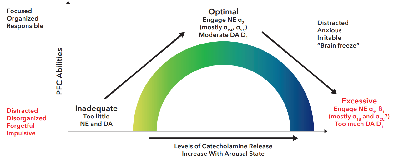
Dysregulation of multiple neurotransmitters is implicated in ADHD, with balanced concentrations of DA and NE required for optimal functioning of the PFC. Low levels of BDNF & Synaptic Plasticity.
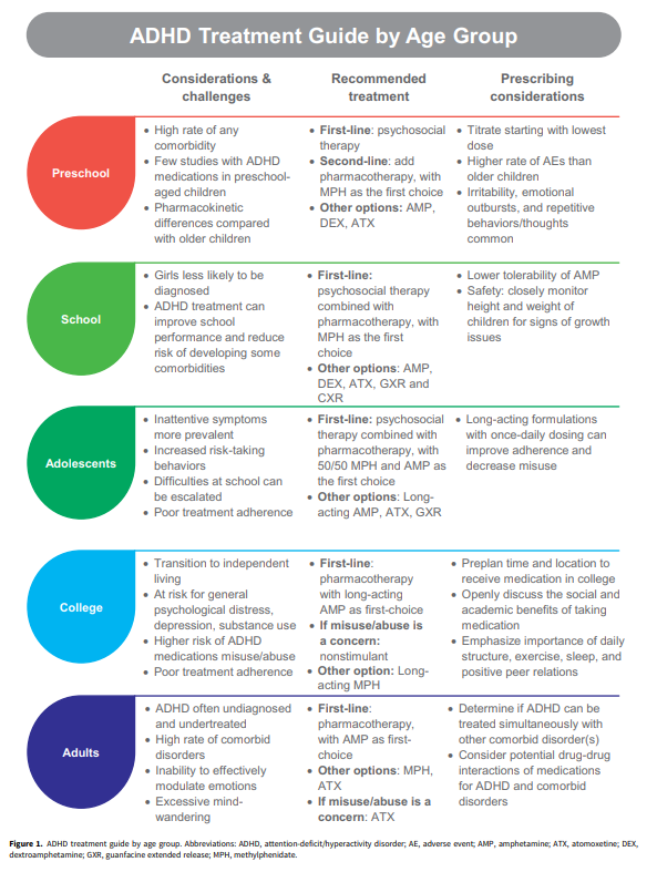
Following diagnosis of ADHD in a child, adolescent or adult, clinicians adopt a multidisciplinary approach as recommended by current treatment guidelines comprising of both non-pharmacological and pharmacological treatment options.
Non-pharmacological therapy includes several procedures as behavioural interventions, such as training of parent behaviour and/or social skills; cognitive
training, focused in reducing ADHD symptoms by improving performance in specific neuropsychological functions using electronic interfaces (computers, tablets, smartphones) or noncomputerized methods that allow performance reassessment so that training is adaptive; neurofeedback based on improving self-control over brain activity patterns (real-time electroencephalogram (EEG) data monitoring of, or coaching programs, focused on improving executive functions.
Pharmacological Treatment for ADHD often involves the prescription of stimulant medications, which paradoxically cause a calming effect in these patients.
Stimulants [amphetamines, lisdexamfetamine dimesylate (LDX), and methylphenidate (MPH)] have generally been used as first-line pharmacologic treatment owing to a higher efficacy in symptomatology reduction compared to non-stimulant medications (atomoxetine)in all groups of age (children, adolescents, and adults) which are considered as second-line medication and are administered when stimulants are contraindicated or because of lack of response or intolerance, in the short-term management of ADHD.
Individualization of attention-deficit/ hyperactivity disorder treatment: pharmacotherapy considerations by age and co-occurring conditions
Individualization of attention-deficit/ hyperactivity disorder treatment: pharmacotherapy considerations by age and co-occurring conditions
Individualization of attention-deficit/ hyperactivity disorder treatment: pharmacotherapy considerations by age and co-occurring conditions